Choosing an implant for Lateral lumbar interbody fusion requires more than comparing cage sizes. The decision affects alignment, stability, bone healing, and the patient’s recovery path. Dr. Luiz Pimenta, a pioneer of the lateral approach, has described it simply: “The lateral approach is a minimally invasive approach to the spine.” That principle matters, but it does not make every implant suitable for every patient.
A reliable selection process begins with the patient’s anatomy. Review standing radiographs, CT scans, and MRI findings together. Measure disc height, segmental lordosis, vertebral endplate shape, and the available corridor through the psoas. A wide cage may improve footprint and reduce subsidence risk. However, it may not fit safely within a narrow disc space. Material also matters. Titanium, porous structures, and polymer options offer different imaging and bone-growth characteristics. Bone density deserves close attention. Fragile endplates can fail under an apparently stable implant.
Small details matter.
The surgeon should also consider whether the cage restores alignment or merely fills the disc space. The ideal implant supports the endplates without excessive distraction. It should match the surgical plan, fixation strategy, and patient-specific risks. No checklist is perfect. Even experienced teams can underestimate endplate weakness or overestimate correction potential. That uncertainty deserves honest discussion. This guide examines implant dimensions, materials, lordotic angles, subsidence resistance, and clinical evidence. It also highlights practical questions surgeons should ask before implantation, while recognizing that final decisions require qualified, patient-specific medical judgment.
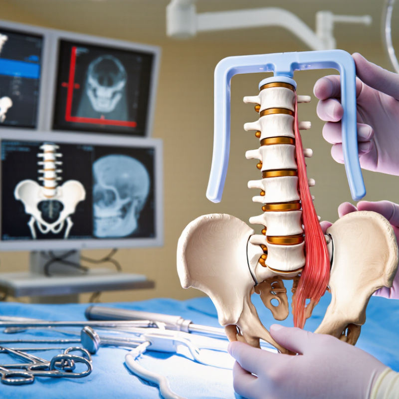
A lateral lumbar interbody fusion implant is a structural spacer placed between lumbar vertebrae. Surgeons insert it through a side approach, usually through the retroperitoneal corridor. Its main purpose is to restore disc height, support the anterior column, and encourage bone growth across the treated segment.
The implant can also improve foraminal height and relieve nerve pressure indirectly. It is not a replacement for careful decompression when severe stenosis remains. Shape, width, height, and material affect stability and alignment. Surgeons must assess endplate strength, bone density, segmental deformity, and the patient’s anatomy before selection. A tall cage may restore height, but excessive distraction can increase pain or endplate injury.
Clinical evidence is encouraging, but not uniform. A systematic review published in the International Journal of Spine Surgery analyzed more than 2,600 patients and reported fusion rates near 90% at 12 months. Results varied by technique, follow-up period, and patient risk factors. That difference matters.
A suitable implant should contact strong peripheral endplate bone. Poor positioning can cause subsidence, especially in osteoporotic vertebrae. Supplemental fixation may improve construct stability in selected cases. However, implant choice alone cannot correct smoking, malnutrition, or poor bone quality. The uncomfortable truth is simple: a technically advanced cage may still fail in the wrong biological environment. Surgeons should combine imaging, bone-density assessment, published outcomes, and hands-on experience. The decision deserves restraint, not guesswork.
Lateral lumbar interbody fusion implant selection begins with the patient, not the cage. Age, activity level, symptoms, and previous surgery shape the surgical objective. Bone density deserves close attention because weak vertebrae may increase subsidence risk. Review CT and MRI together. CT shows endplate strength and bony corridors. MRI clarifies disc height, stenosis, and neural compression. Standing radiographs add another practical detail: sagittal alignment under load.
Anatomy often determines whether a lateral approach is suitable. The surgeon must assess the psoas position, vascular structures, facet joints, and the size of the disc space. A narrow corridor may limit implant width or require a different plan.
Endplate preparation also matters. Excessive removal can weaken support, while inadequate preparation may prevent stable seating. Implant height should restore disc space without creating unnecessary tension on surrounding tissues. Width and footprint should match the available endplate, not simply the largest option.
Patient goals remain important. A younger, active patient may need durable alignment correction, while an older patient may require a more conservative construct. Bone quality, deformity, instability, and the number of treated levels can change fixation needs. No measurement is perfect. Intraoperative findings sometimes challenge the preoperative plan. That is where experience matters, although experience can still be mistaken. Careful imaging review, informed patient discussion, and disciplined surgical judgment should guide the final selection.
How to Choose Lateral Lumbar Interbody Fusion Implants
Implant material influences imaging, bone response, and load transfer. Radiolucent polymers allow clearer postoperative assessment of fusion. Titanium offers strong structural support and may encourage bone integration. Porous surfaces can improve fixation, but they do not eliminate subsidence risk. Material choice should match bone quality, surgical goals, and imaging needs. No material is perfect.
Shape matters as much as material. A wider footprint can distribute force across stronger areas of the vertebral endplate. Anatomical designs may improve contact with the disc space. Angled implants can help restore lumbar alignment when appropriate. However, excessive correction may create unwanted stress. The surgeon should review MRI, CT, and standing radiographs before selecting an implant shape.
Size requires careful judgment. Height should restore disc space without over-distracting the segment. Length and width must fit the vertebral anatomy without breaching the endplate. Larger does not always mean safer. An undersized implant may provide poor support, while an oversized implant can damage bone. In practice, small anatomical differences can change the decision. Even experienced teams may revise their plan after direct intraoperative assessment. That uncertainty deserves attention. Fitness, bone density, previous surgery, and endplate condition all influence the final selection. A reliable choice combines measured anatomy, validated design data, and the surgeon’s clinical experience.
| Selection Dimension | Common Option | Typical Characteristics | Potential Advantages | Important Considerations | When It May Be Appropriate |
|---|---|---|---|---|---|
| Implant Material | Radiolucent PEEK | Polyether ether ketone; radiolucent on CT and radiographs, usually with radiopaque markers to show implant position. | Allows clearer visualization of fusion progression and endplate position; elastic modulus is closer to cortical bone than that of solid metal. | Does not inherently provide a bone-bonding surface. The implant commonly relies on an internal graft chamber and surface design for fusion support. | Useful when postoperative imaging of the graft and fusion mass is a priority. |
| Implant Material | PEEK with a porous or osteoconductive surface | PEEK body combined with a porous coating or surface intended to support bone ongrowth or ingrowth, depending on the design. | Combines radiographic visibility of the PEEK body with a surface intended to improve bone–implant interaction. | Clinical performance depends on pore architecture, coating integrity, graft use, preparation technique, and patient biology. | May be considered when imaging clarity and a more bone-conductive interface are both desired. |
| Implant Material | Titanium alloy | Strong metallic material with high radiodensity; may be machined, textured, or manufactured with a porous structure. | High strength, established surgical use, and compatibility with designs intended to resist deformation and support bone attachment. | Creates more CT artifact than PEEK and may make assessment of the fusion bed less convenient. Solid titanium has a substantially higher stiffness than bone. | May be selected when structural strength, radiographic localization, or a titanium-based fixation interface is important. |
| Implant Material | Porous titanium | Titanium manufactured with interconnected pores or a porous surface designed to encourage bone ingrowth. | Can provide a bone-conductive interface and may reduce the effective stiffness of the implant compared with a fully solid metal design. | Porosity can increase surface roughness and imaging artifact; the implant still requires appropriate endplate preparation and adequate graft biology. | May be considered for patients in whom biological fixation and bone–implant integration are key design goals. |
| Endplate Contact Shape | Rectangular footprint | Broad anterior-to-posterior and medial-to-lateral contact area, commonly used to span a substantial portion of the vertebral endplate. | Broad coverage can improve load distribution and reduce the risk of focal endplate loading when positioned correctly. | Must fit the available disc space and avoid excessive contact with the stronger peripheral rim or malposition relative to the apophyseal ring. | Often considered when endplate coverage and segmental stability are primary objectives. |
| Endplate Contact Shape | Rounded or anatomically contoured footprint | Curved edges or a contour intended to better match the vertebral endplate profile and facilitate insertion through a lateral corridor. | May reduce edge impingement and simplify alignment with the disc space during insertion. | Actual contact area depends on implant position, vertebral anatomy, and the relationship between implant curvature and endplate geometry. | May be useful when the patient’s endplate anatomy or insertion path favors a contoured implant. |
| Sagittal Shape | Parallel implant | Superior and inferior surfaces are approximately parallel, producing little or no built-in segmental lordosis. | Provides a predictable neutral geometry and may be suitable when segmental angulation is already acceptable. | May not provide enough correction in a collapsed or kyphotic disc space; excessive distraction can still occur if height is oversized. | Appropriate when maintaining disc-space height without adding substantial lordotic correction is desired. |
| Sagittal Shape | Lordotic or wedge-shaped implant | Anterior height is greater than posterior height; commonly offered with several nominal lordosis options. | Can help restore segmental lordosis and support correction of selected sagittal alignment problems. | Lordotic correction is affected by implant position, disc preparation, facet condition, and the motion segment’s flexibility. Higher angles may increase endplate loading if poorly matched. | May be considered when restoration of segmental lordosis is part of the surgical plan. |
| Coronal Shape | Low-profile or narrow lateral implant | Smaller medial-to-lateral footprint intended for limited disc spaces or constrained anatomy. | May facilitate insertion when the available corridor or disc-space width is limited. | Smaller footprints generally provide less endplate coverage and may increase the importance of precise placement and endplate preservation. | May be useful when patient anatomy does not accommodate a wider implant. |
| Coronal Shape | Wide lateral implant | Larger medial-to-lateral footprint designed to span more of the vertebral endplate. | Can improve footprint coverage, disc-space stability, and restoration of foraminal height when correctly sized. | Requires sufficient disc-space exposure and careful attention to vascular, psoas, neural, and endplate anatomy. | Often considered when the anatomy permits broad endplate support and indirect decompression is desired. |
| Implant Height | Approximately 8–10 mm | Lower height range used for relatively preserved or moderately collapsed disc spaces, depending on the level and anatomy. | May reduce the risk of over-distraction when the native disc space is not severely collapsed. | Should not be selected solely to maximize foraminal height; excessive height can increase endplate stress and soft-tissue tension. | May fit patients with limited disc-space collapse and adequate residual height. |
| Implant Height | Approximately 10–14 mm | Common mid-range height category for restoring disc-space height in many adult lumbar segments. | Can provide a balance between disc-space restoration, implant seating, and endplate contact when matched to the patient. | Height must be assessed with trial implants and imaging; the same nominal height can have different effects at different lumbar levels. | May be suitable for moderate disc-space collapse when the endplates can safely accommodate the implant. |
| Implant Height | Approximately 14–18 mm or greater | Taller implant category intended for selected severely collapsed or anatomically large disc spaces. | May provide greater disc-space and foraminal-height restoration in appropriately selected cases. | Higher implants can increase distraction, endplate loading, neural or psoas tension, and the risk of subsidence if oversized or poorly positioned. | Reserved for carefully evaluated anatomy with adequate distraction tolerance and preserved endplate strength. |
| Implant Length | Approximately 40–50 mm | Shorter lateral implant length, with the exact choice dependent on vertebral-body dimensions and surgical level. | May be easier to insert through a limited access corridor and can be appropriate for smaller vertebral bodies. | Reduced length may provide less anterior-to-posterior endplate coverage; placement should avoid unsupported or overly peripheral seating. | May be considered when vertebral-body depth or access constraints limit implant length. |
| Implant Length | Approximately 50–60 mm | Longer implant category intended to span a greater portion of the vertebral endplate. | Can increase contact area and improve resistance to migration when the footprint is well matched to the endplate. | Longer is not automatically better; excessive length or anterior placement may create anatomic or vascular concerns. | May be selected for larger vertebral bodies when broad endplate coverage is achievable without unsafe positioning. |
| Graft Chamber | Large central graft window | Open internal chamber designed to hold autograft, allograft, or another approved bone-graft material. | Provides a pathway for bone formation across the disc space and allows a substantial graft volume. | Fusion depends on graft quality, endplate preparation, implant stability, patient factors, and adherence to the device instructions for use. | May be preferred when maximizing graft volume is clinically relevant. |
| Radiographic Markers | Radiopaque marker pins or wires | Markers identify implant boundaries and orientation when the main implant body is radiolucent or partially radiolucent. | Improve assessment of implant position, depth, and alignment during and after surgery. | Markers do not replace intraoperative imaging or direct evaluation of endplate integrity and implant seating. | Important for accurate placement and postoperative monitoring, especially with radiolucent implant bodies. |
| Sizing Principle | Maximum safe endplate coverage without over-distraction | Implant dimensions are selected from imaging, trials, and direct surgical assessment rather than from height alone. | Balances stability and load distribution against the risks associated with an oversized implant. | Oversizing may contribute to endplate damage, subsidence, neurologic symptoms, psoas-related symptoms, or difficult insertion. | Applicable to every LLIF case and should guide final material, shape, height, width, and length selection. |
Choosing a lateral lumbar interbody fusion implant begins with the surgical approach, not the catalog. The access corridor determines implant height, width, and insertion angle. A transpsoas route may require psoas mapping and neuromonitoring. An oblique route can change vascular and retractor considerations. These differences directly affect implant selection. Small anatomical details matter.
Preoperative CT and MRI help assess endplate shape, disc height, bone quality, and foraminal collapse. The implant should match the prepared disc space without excessive distraction. A cage that is too narrow may provide limited support. One that is too tall may increase endplate stress. Fit matters. Compatibility also includes supplemental fixation, graft space, radiographic markers, and insertion instruments. Every component must work within the planned trajectory.
Surgeons should confirm sizing options before positioning the patient. Access can become less forgiving once the table is set. Prior surgery, osteoporosis, deformity, and patient anatomy may change the safest plan. A technically elegant implant is not automatically appropriate. It is easy to overvalue alignment goals and overlook endplate preparation. Careful review of images, surgical steps, and backup options improves reliability. Still, planning is not perfect. Intraoperative findings may require a smaller implant, another fixation strategy, or a different approach. That uncertainty deserves respect.
Choosing a lateral lumbar interbody fusion implant requires more than comparing sizes or cage materials. Safety depends on patient anatomy, bone quality, surgical access, and implant positioning. A narrow corridor near the psoas can increase nerve irritation risk. Preoperative imaging should clarify vascular anatomy, disc height, and endplate strength.
Clinical outcomes need careful interpretation. Pain scores may improve quickly, but early relief does not prove durable fusion. Teams should review fusion rates, subsidence, reoperation frequency, and neurological complications across appropriate follow-up periods. A cage that restores disc height may also overload a fragile endplate. Small details matter. Implant footprint, height, angulation, and graft containment can influence stability over time.
Long-term performance deserves particular attention in osteoporotic patients and multilevel procedures. Follow-up radiographs and, when indicated, computed tomography can reveal settling, migration, or incomplete fusion. No implant removes biological uncertainty. That is an uncomfortable fact. Surgeons must balance published evidence with the individual patient’s risks, goals, and activity level. Evidence may also be limited when studies use short follow-up or inconsistent definitions of fusion. A careful consent discussion should explain these gaps plainly. Even experienced teams can misjudge endplate resistance or underestimate adjacent-segment stress. Continuous outcome tracking can expose those errors and improve future implant selection.
Reported clinical ranges for safety, outcomes, and long-term performance considerations
The chart summarizes commonly reported ranges in peer-reviewed LLIF literature rather than outcomes for a specific implant or manufacturer. Rates vary with patient selection, surgical approach, follow-up duration, cage positioning, supplemental fixation, and study design. Implant selection should consider endplate preservation, subsidence resistance, indirect decompression potential, alignment goals, and compatibility with the patient’s anatomy.