
How to Choose Anterior Lumbar Fusion Solutions
Choosing among Anterior lumbar fusion solutions requires more than comparing implant shapes or promotional claims. The right decision begins with the patient, not the product.
A careful evaluation considers pain patterns, neurological findings, spinal alignment, bone density, previous operations, and vascular anatomy. Imaging should explain the problem clearly. It should not replace clinical judgment. The surgeon must also examine whether anterior access is safe for that specific patient.
Dr. Alexander R. Vaccaro, a respected spine surgeon and researcher, has often emphasized a patient-centered principle: “The best operation is the one that is right for the patient.” That idea deserves practical attention. A technically impressive solution may still be unsuitable for someone with fragile bone, complex anatomy, or unrealistic expectations.
Small details matter. A patient may ask about cage design, graft materials, fixation, recovery time, and the possibility of additional surgery. The surgeon should explain these issues in plain language, including uncertainty. No solution is risk-free.
Not every case needs the same approach.
Experience also matters. Surgeons should understand anterior exposure risks, implant selection, spinal biomechanics, and postoperative rehabilitation. They should discuss evidence rather than promise guaranteed results. Independent verification can help patients compare outcomes, complication rates, and follow-up standards.
This guide examines how to assess Anterior lumbar fusion solutions with a balanced perspective. It will consider clinical goals, surgical access, implant characteristics, patient safety, and long-term expectations. Some choices remain difficult. That is where honest discussion becomes essential.
Understanding Anterior Lumbar Fusion and Its Clinical Purpose
How to Choose Anterior Lumbar Fusion Solutions
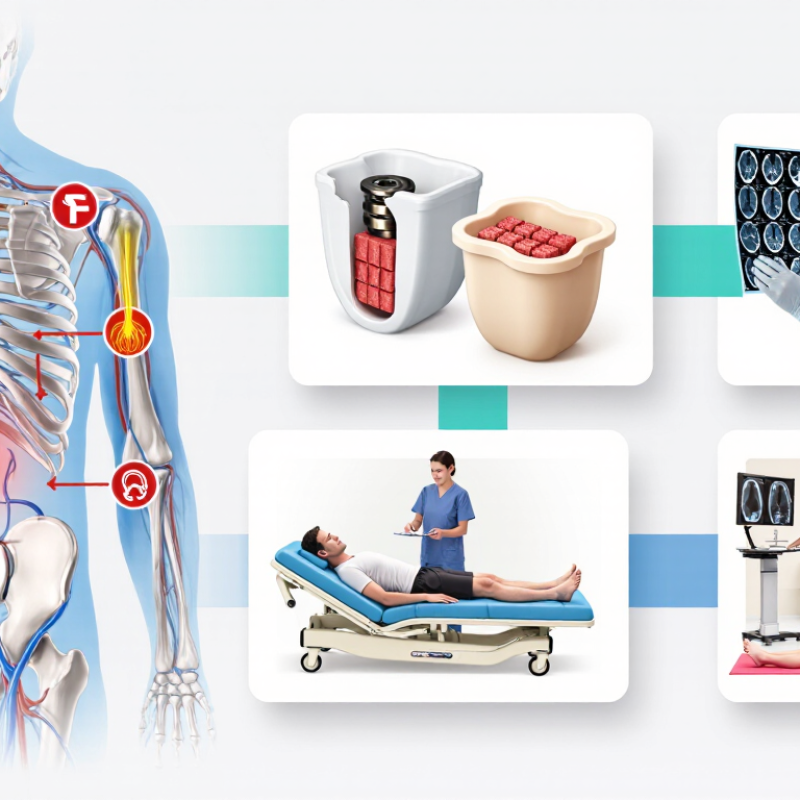
Anterior lumbar fusion is performed through the front of the body to reach the lumbar disc space. The surgeon removes the damaged disc and places a structural spacer with bone graft material. This can restore disc height, improve alignment, and create conditions for bone fusion. The clinical purpose is targeted stabilization, not simply treating every case of back pain.
It may be considered for selected patients with disc degeneration, spinal instability, deformity, or recurrent symptoms after other treatments. Careful assessment comes first. Imaging should be compared with the patient’s symptoms, physical findings, and daily limitations. A scan alone cannot justify surgery. It cannot.
Choosing a solution involves bone quality, disc height, spinal anatomy, prior operations, and the condition of nearby blood vessels. The surgeon should also discuss graft options, fixation needs, healing time, and possible complications. These include infection, nerve symptoms, blood vessel injury, nonunion, and continued pain. Evidence-based planning is important, but it does not replace clinical judgment. Some decisions remain uncertain, especially when symptoms and imaging do not match neatly. A reliable discussion includes alternatives, expected benefits, individual risks, and the surgeon’s experience with the approach. Patient preferences matter too. Pain relief is not guaranteed.
How to Choose Anterior Lumbar Fusion Solutions - Understanding Anterior Lumbar Fusion and Its Clinical Purpose
| Fusion Solution | Primary Access Corridor | Typical Clinical Purpose | Potential Clinical Advantages | Important Risks or Limitations | Key Selection Considerations |
|---|---|---|---|---|---|
| Anterior Lumbar Interbody Fusion (ALIF) | Direct anterior access to the lumbar disc space through the abdomen or retroperitoneal corridor. | Treatment of symptomatic disc degeneration, selected low-grade spondylolisthesis, recurrent disc disease, and restoration of disc height or lumbar alignment. | Provides broad disc-space access, facilitates removal of disc material, allows placement of a large interbody implant, and can support restoration of foraminal height and segmental lordosis. | Vascular injury, retrograde ejaculation in some male patients, ileus, abdominal complications, infection, implant subsidence, nonunion, and approach-related complications. | Evaluate vascular anatomy, prior abdominal surgery, body habitus, osteoporosis, L4–L5 or L5–S1 access, deformity, and the need for direct posterior decompression. |
| Oblique Lumbar Interbody Fusion (OLIF) | Oblique retroperitoneal corridor, generally positioned between the great vessels and the psoas muscle. | Treatment of selected degenerative disc disease, foraminal narrowing, segmental instability, and certain degenerative deformities. | May reduce direct psoas muscle traversal, provides access to a relatively large disc space, and can assist with indirect decompression through restoration of disc and foraminal height. | Vascular or sympathetic-chain injury, ureteral or peritoneal injury, thigh or groin symptoms, inadequate decompression, subsidence, nonunion, and approach difficulty at certain levels. | Review vessel location, psoas anatomy, ureter position, level-specific accessibility, bone quality, stenosis severity, and whether posterior fixation or decompression is required. |
| Lateral Lumbar Interbody Fusion (LLIF) | Direct lateral transpsoas access to the lumbar disc space. | Selected cases of degenerative scoliosis, disc-space collapse, coronal or sagittal imbalance, and foraminal stenosis. | Enables placement of a wide interbody implant, may improve disc and foraminal height, and can provide indirect neural decompression while limiting direct posterior muscle disruption. | Transient or persistent thigh pain, hip-flexor weakness, sensory changes, psoas injury, lumbar plexus injury, vascular or visceral injury, subsidence, and limited access to the lowest lumbar level. | Assess psoas and lumbar plexus anatomy with imaging, determine whether the target level is reachable, and identify fixed stenosis or pathology that may not respond to indirect decompression. |
| Anterior-to-Psoas Interbody Fusion | Anterior or anterolateral retroperitoneal corridor that avoids traversing the psoas muscle when anatomy permits. | Selected lumbar reconstruction procedures requiring disc-height restoration, alignment correction, or indirect decompression. | May decrease direct psoas disruption and permit a relatively broad implant footprint for segmental reconstruction. | Vascular, sympathetic, ureteral, peritoneal, and neural risks remain possible; the corridor can be restricted by vascular anatomy, psoas position, or level-specific structures. | Use detailed cross-sectional imaging to confirm the safe corridor, evaluate the iliac vessels, and determine whether the approach provides adequate access to the intended level. |
| Posterior Interbody Fusion as a Comparator | Posterior midline or paramedian access through the paraspinal muscles and posterior spinal elements. | Direct treatment of central or lateral recess stenosis, instability, spondylolisthesis, and pathology requiring posterior neural decompression. | Provides direct visualization and decompression of neural structures and does not require an anterior or retroperitoneal access corridor. | Potential paraspinal muscle injury, dural tear, epidural scarring, neural injury, blood loss, postoperative pain, and reduced ability to restore anterior column height compared with some anterior approaches. | Consider this option when direct decompression is essential, anterior access is unsuitable, or the pathology is primarily posterior. |
| Hybrid or Staged Reconstruction | Combination of anterior, lateral, and/or posterior corridors performed in one or more operative stages. | Complex deformity, multilevel degeneration, revision surgery, significant alignment loss, or cases requiring both indirect and direct decompression. | Allows the surgical plan to address different anatomical problems through different corridors and may combine alignment correction with targeted neural decompression. | Greater overall procedural complexity, cumulative approach-related risks, longer treatment pathway, and increased need for coordinated perioperative planning. | Assess medical fitness, bone quality, deformity parameters, prior operations, vascular anatomy, neurologic compression, and the risks of single-stage versus staged treatment. |
Related Posts
-

Top 5 Cervical Spine Devices for Effective Treatment Solutions?
-

2026 Best Innovations in Mimetic Metal Technology for Future Applications?
-

China Best Healthcare Procedural Support for Global Buyers?
-
Why Thoracolumbar Implant Systems Are Essential for Spinal Surgery?
-

Top 10 Precision Surgical Instruments for Enhanced Medical Procedures?
-

Best Lumbar Spine Solutions for Global Buyers in China?