Choosing Cervical stabilization technology is a clinical decision, not a simple product comparison. It affects alignment, motion, fixation strength, surgical workflow, and long-term patient safety. The World Health Organization reports that musculoskeletal conditions affect approximately 1.71 billion people worldwide. Neck pain remains a substantial contributor to disability, although reported figures vary by population and diagnostic criteria (WHO, Musculoskeletal Health, 2022). The Global Burden of Disease Study also identifies neck pain as a persistent global health concern. These findings show why technology selection deserves more than marketing claims.
A suitable system should match the patient’s anatomy, pathology, bone quality, and required stability. Surgeons must also examine imaging evidence, implant materials, fatigue performance, fixation design, and clinical outcomes. Regulatory clearance matters, but it does not automatically prove superiority. The U.S. Food and Drug Administration’s 510(k) database can confirm device authorization and substantial equivalence. It cannot replace independent clinical judgment. FDA medical-device reporting data may reveal complications, yet reporting systems have recognized limitations.
The best choice is rarely universal.
Professional guidance from organizations such as AOSpine and the North American Spine Society emphasizes patient selection, evidence quality, and careful postoperative assessment. A 2023 systematic review in the Journal of Neurosurgery: Spine also highlights the importance of comparing radiographic results with patient-reported outcomes. However, real-world results can differ between hospitals, surgeons, and patient groups. This article examines how to evaluate cervical stabilization technology through evidence, experience, and practical risk control. Some decisions remain imperfect. That uncertainty should be acknowledged, not hidden.
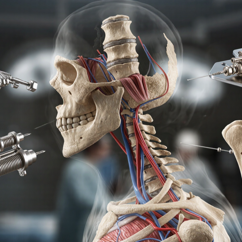
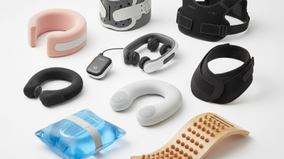
Cervical stabilization technology is designed to limit harmful neck motion while preserving safe, functional movement. Its clinical purpose is not simply to hold the head still. It supports healing, protects vulnerable tissues, and may reduce pain during controlled activity. Clinicians consider instability, fracture patterns, neurological findings, surgical plans, and the patient’s daily environment. A collar, brace, or fixation system must match the problem. More support is not always better.
Choosing technology begins with a careful assessment, not a product comparison. Physical examination and appropriate imaging help clarify alignment, stability, and pressure risks. The fit should distribute contact evenly around the jaw, occiput, and chest. A poorly fitted device can cause skin injury, swallowing difficulty, breathing discomfort, or unwanted movement. Clinicians also check whether the patient can apply, clean, and tolerate it correctly. Small details matter. Age, bone quality, body shape, and communication ability can change the safest option. For postoperative patients, instructions from the surgical team should guide timing and duration.
Evidence supports stabilization when indications are clear, but it does not make every case predictable. Some patients report less pain yet develop stiffness, weakness, or dependence on the device. That tension deserves honest discussion. Follow-up should examine neurological status, skin condition, comfort, alignment, and functional progress. Technology can assist care, but it cannot replace clinical judgment or patient feedback. I would question any recommendation based only on appearance, advertising language, or a single scan.
A clinical comparison of common cervical stabilization approaches and the factors that influence technology selection.
| Technology or construct | Primary clinical purpose | Typical indications | Motion profile | Important selection factors | Common limitations or risks |
|---|---|---|---|---|---|
| External cervical orthosis | Limits cervical movement while the spine or soft tissues heal. | Selected stable injuries, postoperative protection, and short-term symptom control when prescribed by a clinician. | Restricted motion; the degree of control depends on the orthosis design and fit. | Injury stability, required duration, fit, skin condition, airway considerations, and patient compliance. | Pressure injury, discomfort, reduced mobility, swallowing difficulty, and inadequate control if poorly fitted. |
| Anterior cervical plate with interbody fusion | Maintains alignment and supports fusion after removal of a diseased disc or decompression of the spinal cord or nerve root. | Degenerative disc disease, cervical radiculopathy, myelopathy, selected fractures, and instability treated through an anterior approach. | Intended to create a fusion segment with little or no long-term motion at the treated level. | Number of levels, bone quality, alignment, construct length, endplate preparation, and risk of swallowing-related symptoms. | Nonunion, adjacent-segment degeneration over time, dysphagia, implant prominence, and approach-related complications. |
| Posterior screw-and-rod fixation | Provides robust stabilization and alignment control from the posterior side of the cervical spine. | Multilevel instability, traumatic injury, deformity, posterior decompression, tumor, infection, or poor anterior-column support. | Usually reduces motion across the instrumented segments to support fusion. | Extent of instability, vertebral anatomy, vertebral artery position, bone quality, fixation levels, and need for occipital or upper-cervical fixation. | Muscle disruption, wound complications, neurological or vascular injury, hardware failure, and nonunion. |
| Interbody fusion cage without or with supplemental fixation | Restores disc-space height, supports the anterior or posterior column, and provides a space for bone graft to achieve fusion. | Disc-space collapse, foraminal narrowing, degenerative disease, selected trauma, and revision procedures. | Fusion construct; motion at the treated disc space is intentionally eliminated after successful arthrodesis. | Footprint, height, lordotic angle, graft strategy, endplate integrity, subsidence risk, and need for additional fixation. | Subsidence, migration, nonunion, loss of alignment, nerve injury, and inadequate stability when used in unsuitable anatomy. |
| Cervical disc arthroplasty | Decompresses the neural elements while preserving motion at a carefully selected disc level. | Selected one- or two-level symptomatic disc disease with radiculopathy or myelopathy when facet joints, alignment, and bone quality are acceptable. | Designed to preserve segmental motion, although actual motion depends on implant position, anatomy, and healing. | Facet-joint condition, segmental mobility, sagittal alignment, osteoporosis, instability, deformity, and degree of spondylosis. | Heterotopic ossification, wear or migration, implant failure, persistent symptoms, and poor suitability in advanced degeneration or instability. |
| Occipitocervical fixation | Stabilizes the junction between the skull and upper cervical spine when the upper cervical region cannot provide sufficient support. | Craniocervical instability, selected upper-cervical trauma, inflammatory disease, congenital abnormalities, tumor, and complex revision cases. | Substantially restricts motion at the craniovertebral junction and instrumented upper-cervical levels. | Neurological compression, reducibility, bone stock, vertebral artery anatomy, fixation landmarks, and preservation of lower cervical function. | Loss of upper-cervical motion, wound complications, screw malposition, cerebrospinal-fluid leak, nonunion, and neurological or vascular injury. |
| Dynamic or motion-preserving posterior stabilization | Provides controlled support while attempting to retain limited motion in selected segments. | Highly selected cases with instability or deformity in which preservation of some motion is clinically appropriate and evidence supports the specific indication. | Partially motion-preserving; the amount of retained motion varies with design, anatomy, and postoperative healing. | Indication-specific evidence, alignment, facet-joint status, bone quality, implant mechanics, and long-term follow-up requirements. | Limited long-term evidence for some indications, mechanical wear or failure, progression of instability, and possible need for revision. |
Note: Technology selection is patient-specific and should be determined by a qualified spine specialist after clinical examination and appropriate imaging.
Choosing cervical stabilization technology begins with the patient, not the device. Pain location, trauma mechanism, neurological findings, and previous surgery shape the assessment. Ask about hand weakness, numbness, balance changes, and breathing difficulty. These symptoms may indicate spinal cord involvement or changing instability. Short-term comfort is important, but it cannot replace structural protection.
A trained clinician should review appropriate imaging, such as radiographs, CT, or MRI. Each test answers a different question. CT can clarify fractures, while MRI may reveal soft-tissue or cord injury. Dynamic imaging requires caution and should only occur when clinically safe. A collar, brace, or surgical system must match the injury pattern, bone quality, skin condition, and expected activity level. Fit matters. So does tolerance.
Patient needs can change during recovery. Swelling may increase, pain may improve, or neurological signs may appear later. Reassessment is essential. The most stable-looking patient may still have hidden risk. That is an uncomfortable reality. Clinicians should explain restrictions, warning signs, cleaning needs, and follow-up timing in plain language. Compliance is not guaranteed, especially when a device feels restrictive. Better education may prevent avoidable movement. No single scan or measurement provides the whole answer; careful judgment, current evidence, and multidisciplinary review remain necessary.
Cervical stabilization begins with the patient, not the device. Surgeons compare symptoms, imaging, spinal alignment, bone quality, and daily activity.
Anterior cervical discectomy and fusion removes pressure from the nerves and joins neighboring vertebrae. It offers dependable stability, but nearby levels may experience greater stress over time.
Cervical disc replacement preserves motion in selected patients. However, it may not suit severe arthritis, instability, or poor bone quality.
Posterior stabilization can support complex deformities, multilevel disease, or weakened spinal structures. It may involve more muscle disruption and longer recovery.
Tips:
Ask what problem the procedure solves. Request a clear explanation of expected motion, fusion, risks, and revision options. Compare recovery needs with your work, sleep, and hand function. Review imaging with a qualified spine specialist. A second opinion can expose overlooked details.
Technology is only one part of the decision. Surgical experience, patient selection, and follow-up care strongly influence results. A clean scan does not always explain persistent pain. That deserves careful reflection.
Evidence also changes as longer-term studies appear, so impressive early results need context. Patients should discuss swallowing concerns, nerve symptoms, implant durability, and rehabilitation before choosing a method.
The safest choice is usually the one that matches the diagnosis, anatomy, and realistic goals.
Choosing cervical stabilization technology requires more than comparing specifications. Safety begins with patient selection, anatomical fit, and controlled clinical use. A qualified specialist should review imaging, spinal alignment, bone quality, and expected loads. The device should support the intended treatment without restricting necessary movement. Small fitting errors can create pressure points, instability, or discomfort.
Compatibility deserves close attention. Materials should suit the patient’s medical history and the planned procedure. Confirm compatibility with imaging systems, surgical instruments, and related components before treatment. Review cleaning instructions, adjustment limits, and documented testing. Independent evidence matters. Marketing language does not replace clinical data. Look for regulatory authorization, recognized quality systems, and clearly reported performance results. Ask difficult questions.
Long-term performance is harder to judge. Early comfort can hide later loosening, wear, migration, or material degradation. Maintenance access and follow-up requirements should be realistic for the patient. In practice, I would monitor alignment, pain, skin condition, and functional changes over time. A useful design should remain stable during ordinary daily movement. It should also allow timely inspection when symptoms change.
No checklist is perfect. Patient behavior, healing, and surgical technique can alter results. That uncertainty deserves honest discussion. I would be cautious when long-term evidence is limited, even if short-term outcomes appear attractive. Experienced clinicians should compare published data, post-market safety records, and the patient’s actual needs before making a decision.
Evidence-informed comparison of commonly used cervical stabilization approaches across safety, anatomical compatibility, and long-term performance. Scores use a normalized 1–5 scale and are not clinical risk percentages.
Higher scores indicate stronger overall support for the evaluation dimension. Actual results depend on patient anatomy, surgical level, bone quality, alignment goals, implant sizing, and surgical technique. Clinical decisions should be based on peer-reviewed evidence and professional judgment.
Selecting cervical stabilization technology should begin with the patient, not the equipment. A careful assessment includes pain, neurological signs, movement limits, skin condition, and daily activity. A technology may support alignment, reduce unwanted motion, or assist rehabilitation. It should not replace clinical judgment.
Review published evidence and current regulatory requirements before adoption. Ask whether the device fits the intended patient group. Check adjustment ranges, pressure points, cleaning needs, and emergency removal procedures.
Small details matter. A collar that feels secure in the clinic may become uncomfortable after six hours at home. Involve clinicians, therapists, nurses, and the patient during evaluation. Their practical experience can reveal problems that specifications overlook.
Implementation needs a clear protocol. Record the fitting position, wearing schedule, warning signs, and follow-up date. Train staff through demonstrations, not written instructions alone. Confirm that patients can fasten, remove, and inspect the technology safely.
Monitor skin changes, comfort, mobility, and symptom progression. Review these findings regularly. If outcomes remain poor, change the plan rather than defending the original choice. Our first decision may be reasonable, yet still incomplete. Cost, maintenance, staff time, and access to technical support also influence long-term reliability. An effective solution is one that patients can use correctly, clinicians can monitor, and the care team can improve when real-world evidence exposes weaknesses.